
 Italiano
PROTEIN S
|
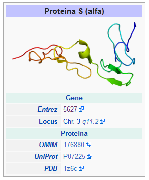
S protein is a vitamin K-dependent protein of 635 amino acids and is primarily synthesized by liver and endothelial cells. It is present in 2 forms: about 40% free (active form) and the remaining 60% is bound to the protein C4BP blood. It has its name in Seattle, the city where it was discovered in 1984.
& Nbsp;
Protein S has the function of protein C cofactor and enhances its activity. It is also very important in maintaining the functionality of retinal photoreceptor cells and phagocytosis of apoptotic cells. In fact, by linking with the phosphatidylserine present in apoptotic cells, the link between the phagocytes and the dying cell is eliminated. This is because protein S binds a TAM receptor on phagocytes acting as a bridge.
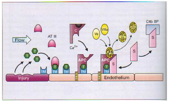
protein S deficiency is a disorder associated with increased risk of venous thrombosis. Protein S, a vitamin-K-dependent physiologic anticoagulant, acts as an enzymatic co-factor of protein C activated in the proteolytic degradation of factor V and factor VIII. Lowering the levels or altering the functionality of protein S involves decreasing Va factor degradation and factor VIII and therefore increasing tendency to venous thrombosis.
There are three types of protein deficiency S hereditary:
- Type I – reduced protein activity S: total protein S decrease (quantitative defect)
- Type II – reduced protein activity S: normal levels of free S protein and total protein levels S are normal (quality defect)
- Type III – Reduced protein activity S: Reduction of protein levels S and normal total protein levels S (quantitative defect)
Protein S deficiency can also be acquired due to deficiency or pharmacological treatment (strains, for example) due to hormone replacement therapy, pregnancy, some liver disease or chronic infections (eg HIV). protein deficit S is the underlying cause of a small percentage of cases of intravascular coagulation (CID), deep venous thrombosis (TVP) and pulmonary embolism (EP).
The schematic representation of the ANTICOAGULANT PROTEIN C / PROTEIN S mechanism is shown below.
| Ref. | Description |
Package |
|||||||
|
COAGULANT
|
|||||||||
| FO220B | PROTEIN S |
40 tests |
|||||||
| Determining the activity of protein S in human plasma. The S protein in the sample is first activated by the addition of a specific serpent venom (Agkistrodon contortrix). Protein S, as a protein co-factor C, inhibits factor V and factor VIII, which involve an elongation of the PTT test, as indicated above: the greater the coagulation time, the greater the amount of S protein present in the sample. The reagent contains the factor Xa and protein C. Lyophilic Reagents. Stability 7 days at -20 ° C.
Reaction at 37 ° C. Dispensing 50 m of plasma lacking S (R2), 50 l of prediluted citrated plasma diluent provided in the kit), 50 l of protein S activator (R1), 50 m < aPTT (R4), 50 calcium chloride (R5). Standard included in the kit (R3). |
|||||||||
|
ELISA
|
|||||||||
| 036-001 | PROTEIN S |
96 test |
|||||||
| Quantitative determination of protein S in human plasma. The S-protein present in the sample binds to monoclonal antibody proteins S human coated on the wells. The plates are then washed to remove unbound proteins and other plasma molecules. Subsequently, a human antibody conjugated with horseradish peroxidase (HRP) is introduced into each well. After incubation, the unbound conjugate is eliminated by washing. The addition of a tetramethylbenzidine (TMB) substrate and hydrogen peroxide (H 2 O 2 ) leads to the development of a blue-colored solution. The addition of sulfuric acid (H 2 SO 4 ) 0.36 N will turn the color of the yellow solution, reducing the TMB and blocking the reaction, denaturing HRP : the optical density measurement at 405 nm is directly proportional to the amount of protein C present in the sample to be examined. The kit does not suffer from interference due to: R506Q mutation Factor V of Leiden, LAC, Factor VIII increase (via PTT) or Activated Factor VII (via PT). Ready-to-use reagents . Stable up to the expiration date.
Reaction at 37 ° C. Pre-diluted and pre-treated sample with polyethylene glycol (PEG), wait 40 minutes for the first incubation, wash the wells, dispense the enzyme conjugate, incubate for 10 minutes, wash the wells, and dispense chromogen. After 10 minutes, block the reaction by dispensing the stop solution. Standard included in the kit (3 bottles, same level). Lyophile to be reconstituted with 0.5 ml of bidistilled water. Stable 24 hours at 2-8 ° C. Do not freeze. |
|||||||||
| 051-001 | Free PROTEIN S |
96 test |
|||||||
| Quantitative determination of free S protein in human plasma. The S-free protein present in the sample binds to monoclonal antibody proteins S free human coated on the wells. The plates are then washed to remove unbound proteins and other plasma molecules. Subsequently, a human free conjugated H-protein peroxidase (HRP) antibody is introduced into each well. After incubation, the unbound conjugate is eliminated by washing. The addition of a tetramethylbenzidine (TMB) substrate and hydrogen peroxide (H 2 O 2 ) leads to the development of a blue-colored solution. The addition of sulfuric acid (H 2 SO 4 ) 0.36 N, will turn the color of the solution to yellow, reducing the TMB and blocking the reaction, denaturing HRP : the optical density measurement at 405 nm is directly proportional to the amount of protein C present in the sample to be examined. The kit does not suffer from interference due to: R506Q mutation V factor Leiden, LAC, factor VIII increase (via the PTT) or activated factor VII (via PT). Liquid ready reagents . Stable up to the expiration date.
Reaction at 37 ° C. Pre-diluted and pre-treated sample with polyethylene glycol (PEG), wait 40 minutes for the first incubation, wash the wells, dispense the enzyme conjugate, incubate for 10 minutes, wash the wells, and dispense chromogen. After 10 minutes, block the reaction by dispensing the stop solution. Standard included in the kit (3 bottles, same level). Lyophile to be reconstituted with 0.5 ml of bidistilled water. Stable 24 hours at 2-8 ° C. Do not freeze. |
|||||||||